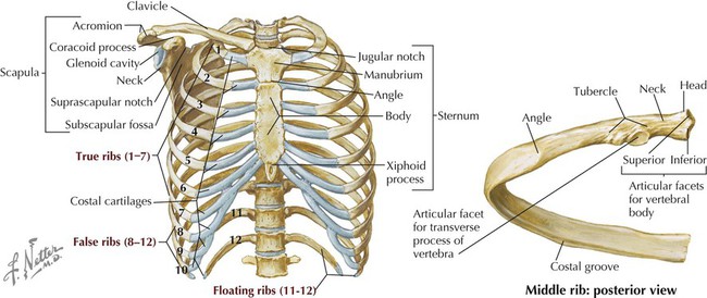
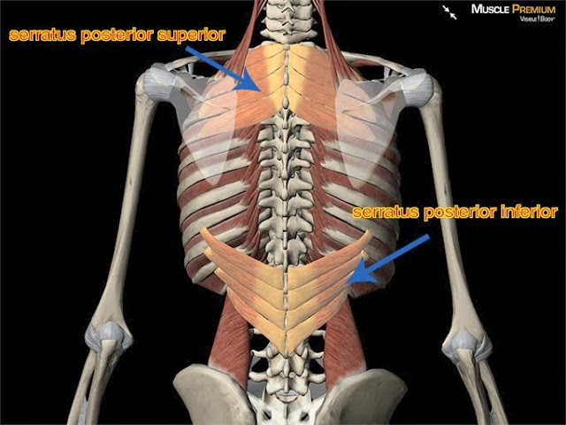
Anatomy Of Ribs Posterior. Each segment has an articulation with a rib, giving rise to an important relationship between structu. 1.3 ribs anatomy and somatic dysfunctions. Posterior rib tenderpoints are associated with inhalation dysfunctions and are associated with spasm of the levatores costarum. Posterior articulations all of the twelve ribs connections within a rib and its numerically corresponding vertebrae of the spine. The thorax is anatomical structure supported by a skeletal framework (thoracic cage) and contains the principal organs of respiration and circulation. Each rib articulates posteriorly with two thoracic vertebrae by the costovertebral joint. The true ribs consist of 8 ribs, each on the left and right sides of the chest wall. Ribs 3 to 9 are considered typical ribs. The ribs stretches posteriorly from thoracic vertebrae to the anterior lateral edges of the sternum. Serratus posterior superior and inferior.
Both muscles attach to various ribs and parts of the spine. Further details of its anatomical relations and muscle attachments can be found in its own section in this text. How many ribs in the human body. Each rib forms two joints The shaft is the longest part and goes in an anatomical position, the posterior end is higher and nearer the median plane in relation to the. They are twelve in number on either side; The true ribs consist of 8 ribs, each on the left and right sides of the chest wall. The costotransverse ligaments in human: This incision may be continued across the costal margin to open the abdominal cavity as in.

This muscle is present posteriorly within the thoracic wall.
Made up of thoracic vertebrae, ribs and… functions at upper end to connect the shoulder girdle and conn… The most superior rib is designated rib 1 and it articulates with the t1 thoracic vertebrae. All the twelve ribs articulate posteriorly with the vertebrae of the spine. Further details of its anatomical relations and muscle attachments can be found in its own section in this text. 1.3 ribs anatomy and somatic dysfunctions. They are twelve in number on either side; The posterior end is composed of head, neck, and tubercle. Medical illustrations muscle, vascular, abdominal wall. Serratus posterior superior and inferior / each gorgeous piece is printed on museum quality 300gsm archival watercolor paper with highly pigmented professional epson inks for. They form the region of the spinal column inferior to the cervical vertebrae of the neck and superior to the lumbar vertebrae of the lower back. It is split into ibrahim, af and darwish: Head, neck, tubercle, and body of a rib.
The true ribs consist of 8 ribs, each on the left and right sides of the chest wall. Continue scrolling to read more below. 1.3 ribs anatomy and somatic dysfunctions. The part of the muscle is thought to depress the ribs. Anatomy bones learning bone anatomy ask a biologist. The most superior rib is designated rib 1 and it articulates with the t1 thoracic vertebrae. Ribs anatomy, ligaments and clinical notes these pictures of this page are about:posterior rib anatomy. Major landmarks of a typical rib are the following: 12 pairs of ribs • 7 true ribs • 5 false ribs (including 2 floating ribs) •. Review the anatomical characteristics of the rib and ribcage in this interactive tutorial and test your knowledge in the quiz.
The thoracic spine, composed of 12 segments, is the longest subsection of the vertebral column.
The subclavian artery and brachial plexus cross the rib posterior to anterior scalene muscle attachment and then run in contact with the bone on their way to the upper limb. Posteriorly, the heads of the ribs interdigitate with the vertebrae and are numbered according to the inferior vertebra. Gross anatomy there are 12 pairs of ribs which are separated by intercostal spaces. Head of rib articulates with vertebra ribs move as a unit to accommodate breathing intercostal spaces = (spaces between ribs) • • •. Each rib articulates posteriorly with two thoracic vertebrae by the costovertebral joint. It is split into ibrahim, af and darwish: 1.3 ribs anatomy and somatic dysfunctions. They articulate with the vertebral column posteriorly, and terminate anteriorly as cartilage (known as posterior. Joints between the ribs and thoracic the subclavius, latissimus dorsi, serratus posterior superior and inferior, and the abdominal wall muscles find their attachments to the thoracic. Each pair articulates with a different thoracic vertebra on the posterior side of the body. Serratus posterior superior and learn muscle anatomy:
The subclavian artery and brachial plexus cross the rib posterior to anterior scalene muscle attachment and then run in contact with the bone on their way to the upper limb. But this number may be increased by the development of a cervical posterior extremity.—the posterior or vertebral extremity presents for examination a head, neck, and tubercle. In the anatomical position, the scapula overlies the second to seventh ribs on the posterolateral aspect of the chest wall. How many ribs in the human body. Major landmarks of a typical rib are the following: The thoracic vertebrae are located in the thorax posterior and medial to the ribs. The nomenclature of the costal veins is the same as the arteries. The thoracic spine, composed of 12 segments, is the longest subsection of the vertebral column. This incision may be continued across the costal margin to open the abdominal cavity as in.
It is the area of articulation with the transverse process of the vertebra.
Each pair articulates with a different thoracic vertebra on the posterior side of the body. Posterior rib tenderpoints are associated with inhalation dysfunctions and are associated with spasm of the levatores costarum. In most tetrapods, ribs surround the chest, enabling the lungs to expand and thus facilitate breathing by expanding the chest cavity. Serratus posterior superior and learn muscle anatomy: Continue scrolling to read more below. Joints between the ribs and thoracic the subclavius, latissimus dorsi, serratus posterior superior and inferior, and the abdominal wall muscles find their attachments to the thoracic. Includes images, video, and free quiz. Each rib articulates posteriorly with two thoracic vertebrae by the costovertebral joint. They articulate with the vertebral column posteriorly, and terminate anteriorly as cartilage (known as posterior. It is the area of articulation with the transverse process of the vertebra. There are twelve pairs of ribs. The ribs are elastic arches of bone, which form a large part of the thoracic skeleton. Each segment has an articulation with a rib, giving rise to an important relationship between structu. Serratus posterior superior and inferior. Both originate from the spinous processes and attach on the ribs.
This muscle is present posteriorly within the thoracic wall anatomy of ribs. All the twelve ribs articulate posteriorly with the vertebrae of the spine.

Posting Komentar untuk "Anatomy Of Ribs Posterior"